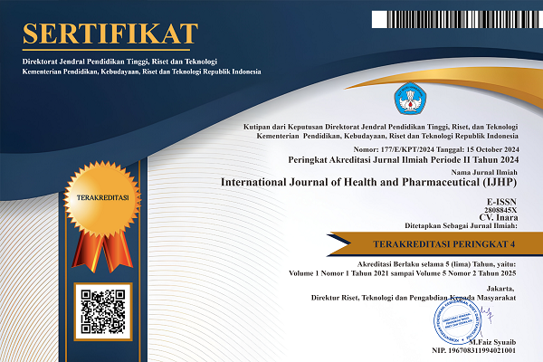
The Effect of Giving Aloe Vera Gel on Collagen Growth in Wistar Rats (Rattus Norvegicus) with Exposure to Ultra Violet-B Light
DOI:
https://doi.org/10.51601/ijhp.v3i4.310Abstract
Exposure to UV rays can cause photochemical damage to the DNA of cells in the body, triggering cancer formation, especially skin cancer in humans. The skin may lose elasticity. Research is needed to ensure that currently, we are still looking for plants or foods that contain collagen, which can prevent wrinkles. This research is an original experiment carried out in a laboratory. Two types of variables play a role in this research: independent variables and dependent variables. In this experiment, aloe vera gel was used as an independent variable as a substitute for UVB light exposure. Collagen development, measured in mouse skin, is the dependent variable here. This study found that the standard control group had no collagen growth and was stable. This was because the typical control treatment was not exposed to UVB rays; hence, collagen formation was stable. The group administered 15% aloe vera extract gel had a more substantial effect on rat skin tissue collagen development after UVB light exposure than the 5% and 10% groups. This is because a higher dose of aloe vera extract includes more chemical components that promote mouse skin collagen formation.
Downloads
References
D. Andrenacci, V. Cavaliere, and G. Lattanzi, “The role of transposable elements activity in aging and their
possible involvement in laminopathic diseases,” Ageing Res. Rev., vol. 57, no. October 2019, p. 100995, 2020,
doi: 10.1016/j.arr.2019.100995.
K. E. Copley and J. Shorter, “Repetitive elements in aging and neurodegeneration,” Trends Genet., vol. 39, no.
, pp. 381–400, 2023, doi: 10.1016/j.tig.2023.02.008.
K. J. Gromkowska-Kępka, A. Puścion-Jakubik, R. Markiewicz-Żukowska, and K. Socha, “The impact of
ultraviolet radiation on skin photoaging — review of in vitro studies,” J. Cosmet. Dermatol., vol. 20, no. 11, pp.
–3431, 2021, doi: 10.1111/jocd.14033.
T. M. Ansary, M. R. Hossain, K. Kamiya, M. Komine, and M. Ohtsuki, “Inflammatory molecules associated
with ultraviolet radiation‐mediated skin aging,” Int. J. Mol. Sci., vol. 22, no. 8, 2021, doi:
3390/ijms22083974.
A. Ali, H. Khan, R. Bahadar, A. Riaz, and M. H. H. Bin Asad, “The impact of airborne pollution and exposure
to solar ultraviolet radiation on skin: mechanistic and physiological insight,” Environ. Sci. Pollut. Res., vol. 27,
no. 23, pp. 28730–28736, 2020, doi: 10.1007/s11356-020-09280-4.
I. Popescu, J. Deelen, M. Illario, and J. Adams, “Challenges in anti-aging medicine–trends in biomarker
discovery and therapeutic interventions for a healthy lifespan,” J. Cell. Mol. Med., vol. 27, no. 18, pp. 2643–
, 2023, doi: 10.1111/jcmm.17912.
A. Vaiserman, A. Koliada, and O. Lushchak, “Phyto-nanotechnology in anti-aging medicine,” Aging (Albany.
NY)., vol. 13, no. 8, pp. 10818–10820, 2021.
E. R. H. Rizza, J. J. DiGiovanna, S. G. Khan, D. Tamura, J. D. Jeskey, and K. H. Kraemer, “Xeroderma
Pigmentosum: A Model for Human Premature Aging,” J. Invest. Dermatol., vol. 141, no. 4, pp. 976–984, 2021,
doi: 10.1016/j.jid.2020.11.012.
F. Schnabel, U. Kornak, and B. Wollnik, “Premature aging disorders: A clinical and genetic compendium,”
Clin. Genet., vol. 99, no. 1, pp. 3–28, 2021, doi: 10.1111/cge.13837.
Z. D. Draelos and A. Shamban, “A pilot study evaluating the anti-aging benefits of a CO2-emitting facial
mask,” J. Cosmet. Dermatol., vol. 22, no. 8, pp. 2198–2204, 2023, doi: 10.1111/jocd.15860.
W. Q. Chng et al., “Topical treatments and skin-resurfacing techniques for skin ageing,” Cochrane Database
Syst. Rev., vol. 2021, no. 8, 2021, doi: 10.1002/14651858.CD014391.
V. Rizzi, J. Gubitosa, P. Fini, and P. Cosma, “Neurocosmetics in skincare-the fascinating world of skin-brain
connection: A review to explore ingredients, commercial products for skin aging, and cosmetic regulation,”
Cosmetics, vol. 8, no. 3, 2021, doi: 10.3390/cosmetics8030066.
P. Newsholme, K. N. Keane, R. Carlessi, and V. Cruzat, “Oxidative stress pathways in pancreatic β-cells and
insulin-sensitive cells and tissues: Importance to cell metabolism, function, and dysfunction,” Am. J. Physiol. -
Cell Physiol., vol. 317, no. 3, pp. C420–C433, 2019, doi: 10.1152/ajpcell.00141.2019.
O. Coskun, M. Kanter, A. Korkmaz, and S. Oter, “Quercetin, a flavonoid antioxidant, prevents and protects
streptozotocin-induced oxidative stress and β-cell damage in rat pancreas,” Pharmacol. Res., vol. 51, no. 2, pp.
–123, 2005, doi: 10.1016/j.phrs.2004.06.002.
J. M. Powers and J. E. J. Murphy, “Sunlight radiation as a villain and hero: 60 years of illuminating research,”
Int. J. Radiat. Biol., vol. 95, no. 7, pp. 1043–1049, 2019, doi: 10.1080/09553002.2019.1627440.
M. Wacker and M. F. Holick, “Sunlight and Vitamin D: A global perspective for health,” Dermatoendocrinol.,
vol. 5, no. 1, pp. 51–108, 2013, doi: 10.4161/derm.24494.
A. Juzeniene and J. Moan, “Beneficial effects of UV radiation other than via vitamin D production,”
Dermatoendocrinol., vol. 4, no. 2, pp. 109–117, 2012, doi: 10.4161/derm.20013.
K. Wake, K. Sasaki, and S. Watanabe, “Conductivities of epidermis, dermis, and subcutaneous tissue at
intermediate frequencies,” Phys. Med. Biol., vol. 61, no. 12, pp. 4376–4389, 2016, doi: 10.1088/0031-
/61/12/4376.
International Journal of Health and Pharmaceutical
R. N. Rohmah, “Microneedling dengan Platelet-rich Plasma (PRP) untuk Skar Akne Atrofi,” Cermin Dunia
Kedokt., vol. 48, no. 9, p. 380, 2021, doi: 10.55175/cdk.v48i9.1500.
E. Bhatia, D. Kumari, S. Sharma, N. Ahamad, and R. Banerjee, “Nanoparticle platforms for dermal antiaging
technologies: Insights in cellular and molecular mechanisms,” Wiley Interdiscip. Rev. Nanomedicine
Nanobiotechnology, vol. 14, no. 2, pp. 1–31, 2022, doi: 10.1002/wnan.1746.
A. Bielach-Bazyluk et al., “Sirtuin 1 and skin: Implications in intrinsic and extrinsic aging—a systematic
review,” Cells, vol. 10, no. 4, pp. 1–21, 2021, doi: 10.3390/cells10040813.
S. Qiu et al., “Hindered phenolic antioxidant passivation of black phosphorus affords air stability and free
radical quenching,” J. Colloid Interface Sci., vol. 606, pp. 1395–1409, 2022, doi: 10.1016/j.jcis.2021.08.098.
C. N. Ginting, I. N. E. Lister, E. Girsang, D. Riastawati, H. S. W. Kusuma, and W. Widowati, “Antioxidant
Activities of Ficus elastica Leaves Ethanol Extract and Its Compounds,” Mol. Cell. Biomed. Sci., vol. 4, no. 1,
p. 27, 2020, doi: 10.21705/mcbs.v4i1.86.
D. Hekmatpou, F. Mehrabi, K. Rahzani, and A. Aminiyan, “The effect of aloe vera clinical trials on prevention
and healing of skin wound: A systematic review,” Iran. J. Med. Sci., vol. 44, no. 1, pp. 1–9, 2019.
T. Chakraborty, S. Gupta, A. Nair, S. Chauhan, and V. Saini, “Wound healing potential of insulin-loaded
nanoemulsion with Aloe vera gel in diabetic rats,” J. Drug Deliv. Sci. Technol., vol. 64, no. April, p. 102601,
, doi: 10.1016/j.jddst.2021.102601.
N. Deora and K. Venkatraman, “Aloe vera in diabetic dyslipidemia: Improving blood glucose and lipoprotein
levels in pre-clinical and clinical studies,” J. Ayurveda Integr. Med., vol. 13, no. 4, p. 100675, 2022, doi:
1016/j.jaim.2022.100675.
F. Haghani, M. R. Arabnezhad, S. Mohammadi, and A. Ghaffarian-Bahraman, “Aloe vera and StreptozotocinInduced Diabetes Mellitus,” Rev. Bras. Farmacogn., vol. 32, no. 2, pp. 174–187, 2022, doi: 10.1007/s43450-
-00231-3.
N. Rohmawati, “Efek Penyembuhan Luka Bakar Dalam Sediaan Gel Ekstrak Etanol 70 % Daun Lidah Buaya
( Aloe Vera L .) Pada Kulit Punggung Kelinci New Zealand Nina Rohmawati K 100040151 Fakultas Farmasi,”
Universitas Muhammadiyah Surakarta, 2008.
M. N. zadeh Gharaboghaz, M. R. Farahpour, and S. Saghaie, “Topical co-administration of Teucrium polium
hydroethanolic extract and Aloe vera gel triggered wound healing by accelerating cell proliferation in diabetic
mouse model,” Biomed. Pharmacother., vol. 127, no. April 2019, p. 110189, 2020, doi:
1016/j.biopha.2020.110189.
S. Notoatmodjo, Metodologi Penelitian Kesehatan, 3rd ed. Jakarta: Rineka Cipta, 2018.
B. Suwarno and A. Nugroho, Kumpulan Variabel-Variabel Penelitian Manajemen Pemasaran (Definisi &
Artikel Publikasi), 1st ed. Bogor: Halaman Moeka Publishing, 2023.
L. C. Boudghane et al., “Phytochemical, antioxidant, and antimicrobial attributes of different extracts of seeds:
the Algerian variety of dates ‘Deglet Nour’ (Phoenix dactylifera L.),” Vegetos, vol. 36, no. 2, pp. 559–565,
, doi: 10.1007/s42535-022-00413-3.
S. H. Ismail, A. Hamdy, T. A. Ismail, H. H. Mahboub, W. H. Mahmoud, and W. M. Daoush, “Synthesis and
characterization of antibacterial carbopol/ZnO hybrid nanoparticles Gel,” Cryst. MDPI, vol. 11, no. 9, pp. 1–23,
, doi: 10.3390/cryst11091092.
E. Mawarni, C. N. Ginting, L. Chiuman, E. Girsang, R. A. S. Handayani, and W. Widowati, “Antioxidant and
Elastase Inhibitor Potential of Petals and Receptacle of Rose Flower (Rosa damascena),” Pharm. Sci. Res., vol.
, no. 2, pp. 105–113, 2020, doi: 10.7454/psr.v7i2.1016.
S. S. Virani et al., Heart disease and stroke statistics—2020 update a report from the American Heart
Association, vol. 141, no. 9. 2020. doi: 10.1161/CIR.0000000000000757.

Downloads
Published
How to Cite
Issue
Section
License
Copyright (c) 2023 Inola Fitria , Djamin Djamin, Buter Samin

This work is licensed under a Creative Commons Attribution-NonCommercial 4.0 International License.